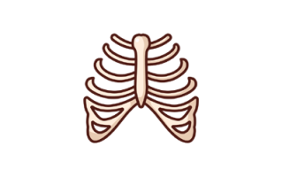
The Intercostal Space is between two adjacent ribs, with 11 intercostal spaces on each side. It consists of intercostal muscles and a neurovascular bundle, with the vein being the most superior structure. The space is separated from the underlying pleura by loose connective tissue.
Download Intercostal Space PPT
| File Name | Intercostal-Space.pptx |
| File Size | 4.93 MB |
| Number of Slides | 39 |
| PPT Preview | Video |
| Author | mbbsppt.com |
| Built on | Microsoft Office 2021 |
| Font used | Gill Sans MT |
Intercostal Space PPT Summary
- The intercostal space is crucial for the thoracic wall’s structure and function.
- Understanding the anatomy and contents of the intercostal space is essential for medical procedures and conditions.
- Differentiation between typical and atypical intercostal spaces is based on blood and nerve supply.
- Intercostal arteries and veins play a vital role in supplying the thoracic wall.
- Procedures like intercostal nerve blocks and thoracotomy involve the intercostal space.
Readme:
- Distribute: You are allowed to use and distribute the presentation for free. If you are willing to share the above presentation, use the page URL to share it anywhere.
- Share your PPT: If you are a doctor/student and want to share your Presentation (PPT) on mbbsppt.com, use this form to upload it. Once you have submitted the presentation, please allow us to publish it in 1-2 days.
- Credits: The above Intercostal Space PPT (presentation) is created by mbbsppt.com. We reserved the right to modify and distribute the presentation.